Osteoporosis Diagnosis: Tests and Methods for Early Detection
Osteoporosis is a condition that causes bones to become weak and brittle, significantly increasing the risk of fractures. Because osteoporosis often progresses without symptoms, early detection is crucial to prevent complications. With the right diagnostic tools, healthcare providers can assess bone health and take necessary steps to strengthen bones before severe damage occurs. In this article, we will explore the various tests and methods used for early detection of osteoporosis, providing insights into how the condition is diagnosed.
Why Early Detection is Important
Osteoporosis is often called the “silent disease” because it progresses gradually and doesn’t show obvious symptoms until a bone fracture occurs. The most common fractures related to osteoporosis affect the hip, spine, and wrist, and these fractures can severely impact a person’s quality of life. Identifying osteoporosis early allows for timely interventions, such as lifestyle changes, dietary modifications, or medications, that can help maintain or even increase bone density, ultimately reducing fracture risk.
Who Should Get Tested for Osteoporosis?
Certain individuals are at higher risk of developing osteoporosis and should consider early screening. The following groups are particularly encouraged to undergo testing:
- Postmenopausal Women: The decline in estrogen levels after menopause can lead to accelerated bone loss.
- Men Over 70: Older men can also develop osteoporosis, especially if they have other risk factors like low testosterone or a history of fractures.
- Individuals with a Family History: If a close family member has had osteoporosis or related fractures, the risk is higher.
- People with Risk Factors: Those with conditions like hyperthyroidism, chronic steroid use, smoking, or excessive alcohol consumption are at an increased risk.
- Individuals with Low Body Weight: Thin or small-framed individuals have less bone mass to begin with, putting them at higher risk for osteoporosis.
Diagnostic Tests for Osteoporosis
Several diagnostic tests are available to evaluate bone health and detect osteoporosis. Each test plays a unique role in determining bone mineral density and assessing fracture risk.
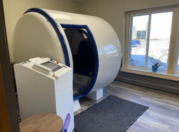
1. Dual-Energy X-ray Absorptiometry (DEXA) Scan
The Gold Standard of Osteoporosis Diagnosis
The DEXA scan is considered the most reliable and commonly used test for diagnosing osteoporosis. It measures bone mineral density (BMD) at key sites, such as the hip and lumbar spine, where fractures are most likely to occur. The DEXA scan is non-invasive, uses minimal radiation, and provides an accurate assessment of bone density.
How It Works: During a DEXA scan, a patient lies on a table while a low-dose X-ray machine measures the density of their bones. The results are provided as a T-score, which indicates how much bone mass differs from that of a healthy young adult:
Normal: T-score of -1.0 or above
Osteopenia (Low Bone Mass): T-score between -1.0 and -2.5
Osteoporosis: T-score of -2.5 or lower
The DEXA scan is a quick and painless procedure that typically takes about 10-15 minutes, providing an essential benchmark for tracking bone density changes over time.
2. Quantitative Ultrasound (QUS)
A Convenient, Radiation-Free Option
Quantitative ultrasound is a portable, radiation-free tool that can be used to assess bone density, usually at the heel (calcaneus). It measures the speed of sound waves passing through bone tissue, which provides an indication of bone strength and elasticity. While it is not as precise as a DEXA scan, QUS is a useful initial screening tool, particularly in settings where access to more advanced imaging is limited.
How It Works: The foot is placed in an ultrasound machine, and sound waves are transmitted through the heel bone. The resulting score can help determine whether further testing, such as a DEXA scan, is needed.
3. Peripheral Dual-Energy X-ray Absorptiometry (pDEXA)
Assessing Bone Density in Peripheral Bones
pDEXA is a type of bone density test that is used to measure bone density in peripheral bones, such as the wrist, finger, or heel. Like the full-body DEXA scan, pDEXA uses low-dose X-rays to evaluate bone mineral density but is generally used as a screening tool rather than for diagnostic purposes.
How It Works: A small, portable X-ray machine is used to measure bone density in peripheral sites. If the test indicates low bone density, further testing at central sites (hip and spine) with a full-body DEXA scan is recommended.
4. Quantitative Computed Tomography (QCT)
3D Assessment of Bone Density
Quantitative computed tomography is a more advanced imaging technique that provides a 3D image of bone, allowing for a detailed assessment of both cortical (outer layer) and trabecular (spongy inner layer) bone. QCT is particularly useful for evaluating bone strength in the spine. It involves more radiation exposure compared to DEXA but provides a detailed assessment of bone quality.
How It Works: A CT scanner takes cross-sectional images of the bones, which are then used to calculate bone mineral density. The resulting images provide a more complete picture of bone structure compared to other methods.
5. Bone Turnover Markers
Assessing Bone Metabolism
Bone turnover markers are substances found in the blood or urine that reflect the rate of bone formation or resorption. While these markers cannot diagnose osteoporosis on their own, they can provide valuable information about bone metabolism. They are often used alongside bone density measurements to assess how quickly a person is losing bone and to evaluate how well they are responding to treatment.
How It Works: Blood or urine samples are tested for specific markers such as C-terminal telopeptide (CTX), which indicates bone resorption, or procollagen type 1 N-terminal propeptide (P1NP), which reflects bone formation. These markers can help determine whether osteoporosis treatments are effective in reducing bone turnover.
Fracture Risk Assessment Tools
In addition to bone density testing, tools like the FRAX (Fracture Risk Assessment Tool) are used to estimate an individual’s 10-year risk of fractures. This tool takes into account multiple factors, including age, gender, weight, history of fractures, and lifestyle habits. The FRAX tool is often used in combination with DEXA scan results to determine whether medication or other treatments are necessary.
What to Do if You Are Diagnosed with Osteoporosis
If a bone density test indicates osteoporosis or osteopenia, there are several steps you can take to reduce bone loss and prevent fractures:
- Medications: There are several types of medications available for treating osteoporosis, including bisphosphonates, hormone replacement therapy (HRT), and selective estrogen receptor modulators (SERMs). These medications help increase bone density and reduce the risk of fractures.
- Nutrition: Adequate intake of calcium and vitamin D is essential for bone health. Dairy products, leafy greens, and fortified foods are good sources of calcium, while sunlight exposure and fatty fish can help boost vitamin D levels.
- Exercise: Weight-bearing exercises, such as walking, jogging, and resistance training, help stimulate bone growth. Exercises that improve balance and coordination, such as tai chi or yoga, can also help prevent falls.
- Lifestyle Changes: Avoid smoking and limit alcohol intake, as both can contribute to bone loss. It’s also important to maintain a healthy weight, as being underweight can increase the risk of fractures.
Conclusion
Early detection of osteoporosis is crucial for preventing fractures and maintaining a good quality of life. With diagnostic tools like the DEXA scan, quantitative ultrasound, and bone turnover markers, healthcare providers can effectively assess bone health and detect osteoporosis before serious complications occur. If you are at risk for osteoporosis, talk to your healthcare provider about undergoing bone density testing. Early diagnosis, along with lifestyle modifications and medical interventions, can help preserve bone strength and reduce the risk of fractures, ensuring a more active and healthy future.